
The Silent Threat of Polycystic Kidney Disease: Early Detection and MRI Screening
Polycystic Kidney Disease (PKD) is an inherited disorder in which fluid-filled cysts develop and gradually replace healthy kidney tissue. Because the kidneys compensate for a long time, many people feel entirely well while the disease is already advancing. By the time symptoms appear or blood tests turn abnormal, significant kidney tissue may already be lost. For families affected by PKD, finding the disease early — and knowing how quickly it is likely to progress — changes what can be done about it.
What is Polycystic Kidney Disease (PKD)?
PKD is a progressive hereditary condition. Its most common form, Autosomal Dominant Polycystic Kidney Disease (ADPKD), affects roughly 1 in every 1,000 people worldwide and is the leading inherited cause of kidney failure. A child of an affected parent has a 50% chance of inheriting the condition.
Left unmonitored, the continued growth of kidney cysts can lead to hypertension, chronic kidney disease, and eventually end-stage renal disease requiring dialysis or kidney transplantation.
Common Symptoms of PKD Include:
-
High blood pressure (hypertension)
-
Back or side (flank) pain
-
Blood in the urine (hematuria)
-
Recurrent urinary tract infections (UTIs) or kidney stones
-
Abdominal swelling or a feeling of fullness
Why Blood Tests Can Miss Early PKD
Standard blood tests — serum creatinine and estimated glomerular filtration rate (eGFR) — often remain normal in early PKD. The remaining healthy kidney tissue increases its workload to compensate for areas damaged by cysts, holding filtration values within the normal range even as the kidneys double or triple in size.
A normal eGFR is therefore reassuring about kidney function today, but it reveals very little about how much cyst burden has accumulated or how fast it is growing. Imaging shows that structural change directly, often years before filtration begins to decline.
What is MRI Polycystic Kidney Screening and Evaluation?
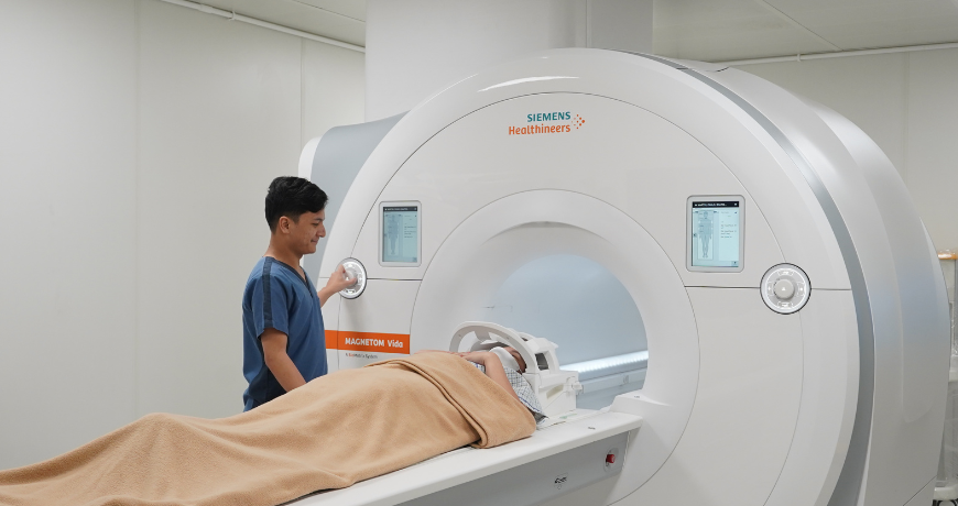
MRI Polycystic Kidney Screening and Evaluation at St. Luke’s Medical Center is a dedicated, non-invasive kidney MRI protocol that accomplishes two things in a single examination:
-
Detection. High-resolution MRI resolves small, subcentimeter cysts and defines their number and distribution in detail, supporting confident assessment in at-risk individuals — including younger adults, in whom cysts are still few and small.
-
Risk stratification. The same examination measures Total Kidney Volume, allowing nephrologists to estimate how rapidly the disease is likely to progress.
Detection answers whether cysts are present. Risk stratification answers the question patients and their nephrologists actually need to act on: how fast is this moving, and what should we do now?
Measuring Disease Severity: TKV and htTKV
Total Kidney Volume (TKV) is the most extensively studied imaging measure in ADPKD. It has been formally qualified by the U.S. Food and Drug Administration as a prognostic biomarker for the disease — a designation reserved for measurements with strong evidence linking them to future outcomes. Rather than judging severity from the appearance of cysts alone, TKV quantifies the actual volume of both kidneys.
Adjusted for the patient’s height (height-adjusted TKV, or htTKV) and interpreted against their age, this measurement places the patient within a validated imaging classification that separates slowly progressive from rapidly progressive disease. Repeat examinations then establish the annual growth rate, producing a measured trajectory rather than an estimate.
That distinction is what determines how closely a patient needs to be followed, and whether they may benefit from disease-modifying therapies.
No Contrast. No Radiation. No Injection.
The examination is performed without contrast dye and without any radiation exposure. There is no intravenous line, no iodinated contrast, and no gadolinium. The sequences used to measure kidney volume rely on the natural signal of the fluid within the cysts, which MRI depicts in high contrast on its own.
For patients screened because of family history, this matters a great deal. Monitoring PKD means imaging repeatedly over decades — and a protocol requiring neither radiation nor an injection can be safely repeated for a lifetime, including in young adults and in patients whose kidney function is already reduced.
The study is performed on an outpatient basis and asks only that the patient lie still and follow simple breathing instructions.
Who Should Consider MRI Polycystic Kidney Screening and Evaluation
-
Adults with a parent, sibling, or child diagnosed with polycystic kidney disease
-
Individuals with unexplained high blood pressure at a young age
-
Patients with a suspected or previously identified hereditary kidney disorder
-
Patients with known PKD who need a baseline or follow-up kidney volume measurement to guide management
-
Prospective living kidney donors from families affected by PKD
Before You Schedule: What Your Nephrologist Will Discuss
Because PKD is inherited, the result of this examination carries information not only for you but for your immediate family. For that reason, MRI Polycystic Kidney Screening and Evaluation is arranged through a consultation rather than booked as a stand-alone scan.
Before imaging, your St. Luke’s nephrologist will discuss:
-
what a positive or a negative result would mean at your particular age
-
what the findings could imply for your parents, siblings, and children
-
how a confirmed diagnosis may affect insurance or HMO coverage and future applications
-
what would change in your care if cysts are found — blood pressure targets, lifestyle measures, monitoring intervals, and eligibility for disease-modifying therapies
-
considerations around family planning
For children and adolescents who have a family history but no symptoms and normal blood pressure, testing is generally deferred until adulthood, so the individual can make an informed decision for themselves.
Most patients find this conversation valuable in itself. Understanding what a result would mean before you have it is what turns that result into something you can act on.
Key Benefits of MRI Polycystic Kidney Screening and Evaluation
-
Earlier detection. Identifies subcentimeter cysts and characterizes cyst burden before blood tests register any change.
-
Objective baseline. Establishes an accurate, reproducible kidney volume against which future growth can be measured.
-
Prognostic clarity. Places patients on a validated progression scale, distinguishing those who need close follow-up from those who do not.
-
Timely intervention. Enables blood pressure control, lifestyle modification, and consideration of disease-modifying therapies during the window when they are most effective.
-
Safely repeatable. No radiation and no contrast, so monitoring can continue for as long as it is needed.
Schedule a Polycystic Kidney Disease Evaluation
If you have a family history of kidney disease, or you want to understand your renal health more clearly, a consultation with a St. Luke’s nephrologist can determine whether MRI Polycystic Kidney Screening and Evaluation is appropriate for you.
For more information about St. Luke’s Medical Center and its services, call our Product Information Hub at 09479930471 / 09399161805 / 09285200239 / 09985822276 or email productinfo@stlukes.com.ph.


