Dynamic Bracing in Scoliosis Rehabilitation
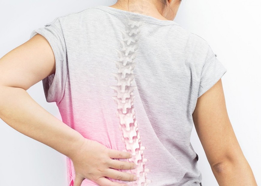
The Mayo Clinic defines Scoliosis as a sideways curvature with rotation of the spine that occurs most often during the growth spurt just before puberty. While scoliosis can be caused by conditions such as cerebral palsy and muscular dystrophy, the cause of most scoliosis is unknown. Studies have now indicated that most forms of Idiopathic Scoliosis is due to improper genetic expression during growth or spinal development.
Most cases of scoliosis are mild, but some children develop spine deformities that continue to get more severe as they grow. Severe scoliosis can be disabling. It can be seen as a C or S curve or increased hunching. An especially severe spinal curve can reduce the amount of space within the chest, making it difficult for the lungs to function properly. It can also lead to growth disharmony, neuromusculoskeletal dysfunction, and functional and structural interference to the nervous system.
Those who have mild scoliosis are monitored closely, usually with X-rays, to see if the curve is getting worse. In many cases, no treatment is necessary.
Some children will need to wear a brace to stop the curve from worsening. Others may need surgery to keep the scoliosis from worsening and to straighten severe cases of scoliosis.
Full-time rigid scoliosis bracing can be an outdated treatment. Full-time bracing often causes more problems for the person wearing it, such as pain that didn't exist before, breathing problems, and weakened muscles. It hasn't been consistently proven to prevent scoliosis surgery, either.
Braces like the most common thoracolumbar-sacral-orthosis brace (TLSO) brace squeeze the chest wall and abdomen. A Norwegian study of the TLSO found it significantly decreases pulmonary functions, including breathing capacity, oxygen, and CO2 exchange ratios. Breathing impacts hormone regulation, muscle and fat composition, and cognitive performance. One study from the University of Athens, Greece showed that children who wore a hard brace had a 30 percent decrease in vital capacity (VC) and a 45 percent decrease in expiratory reserve volume (the air you can push out after a normal exhale). Respiratory distress causes headaches, anxiety, sleep disturbances, nightmares, and cognitive dysfunction (memory, perception, and problem solving).
Primarily, the SpineCor bracing method is an adjustable, non-invasive technique that provides flexible, inconspicuous correction that continues as a child moves and grows. Unlike traditional rigid systems, the SpineCor brace consists of four major components: (1) a plastic pelvic base, (2) a cotton bolero or vest, (3) tie bands and (4) four adjustable or “dynamic” bands. This unique combination of components is simple to use, comfortable to wear, and most importantly, effective in its results. The goal of the dynamic brace is to maintain and improve spinal deformity while re-educating the body to return to a more normal posture.
The SpineCor Dynamic Corrective Brace is a flexible brace that is principally prescribed for Idiopathic scoliosis patients with a Cobb angle between 15° and 50°, Risser sign 0 to 3 or pre-menarche.
The brace was invented by a team of 65 researchers led by Professor Charles H Rivard M.D. and Christine Coillard M.D. of the Research Center, Sainte-Justine Hospital (Montreal, Quebec, Canada). It took more than 12 years and millions of dollars in research funding to develop the world's first truly dynamic corrective brace for Adolescent Idiopathic Scoliosis.
Initial brace fitting and follow-up treatments are supported by the clinical diagnostic software, SpineCor Assistant Software (SAS).
The treatment approach by SpineCor brace is to bring about global postural re-education and progressive correction of curve over time. Unlike rigid brace which "forces" correction, SpineCor brace does not deliver drastic curve correction initially. However, the correction or stabilization gained by wearing the dynamic brace over time is more stable with lower rate of curve progression post-brace. It is able to stop 77% of cases leading to surgery. It was able to achieve 95.7 % stabilization of scoliosis even after removing and not wearing the brace for 2 years. It is also 3.9 times more effective than other bracing procedures. It creates dynamic spinal off-loading and neuromuscular rehabilittaion with the ultimate goal of neuromuscular integration.
The brace is prescribed to be worn by the patients 20 out of 24 hours per day until they have reached skeletal maturity. Radiological evaluations are performed prior to and immediately following the fitting of the brace, and every 4 to 6 months afterwards. To accommodate for growth and postural changes, corrective bands need to be adjusted frequently and require replacement each 6-12 months for optimum brace performance. Major brace components can last from 1.5 - 2 years.
While there are guidelines for mild, moderate and severe curves, the decision to begin treatment is always made on an individual basis according to the Mayo Clinic.
The following will be performed:
- Clinical Evaluation
- X-ray Evaluation
- Posture Evaluation
Not all physicians are currently equipped to treat patients with the SpineCor brace, but information is readily available to qualified practitioners who routinely diagnose idiopathic scoliosis. The Practitioner may use the digital imaging system and assistant software (SAS ) in their office to take the initial body measurements and to arrange for follow-up visits. The brace will then be fitted, and the patient is taught how to use it effectively. Generally, to achieve maximum results, the brace should be worn during the day and may be worn for up to 20 hours at a time. Physical Therapy will offer a program for improved body mechanics and exercises while wearing the brace. Corrective movements gently guide posture and spinal alignment in an optimal direction.
The St. Luke’s Department of Physical Medicine and Rehabilitation in coordination with SpineCor will have a training program for practitioners this February 1 in St. Luke’s-Quezon City and February 2 at St. Luke’s-Global City. A Public seminar regarding this new brace will also be held at St. Luke’s-Quezon City on February 1 and at St. Luke’s -Global City on February2 from 5:00 PM -7:00 PM.
Dr. Reynaldo R. Rey-Matias is the Head of the Department of Physical Medicine and Rehabilitation in both St. Luke’s Medical Center-Quezon City and Global City. He is currently the Vice President of the Asian Oceanian Society of Physical and Rehabilitation Medicine (AOSPRM ). He is a past President of the ASEAN Rehabilitation Medicine Association (ARMA) and the Philippine Academy of Rehabilitation Medicine (PARM) and a past Chairman of the Philippine Board of Physical and Occupational Therapy of the Professional Regulation Commission.


